
medical film 35*35
1,040.00
Out of Stock
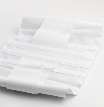
Medical Film 35*35 refers to a square-sized radiographic film that measures 35 cm by 35 cm. This size is commonly used in medical imaging procedures when a large and balanced view of a specific body part is needed. It is typically used for imaging areas such as the chest, spine, or other larger anatomical structures, where a square format is preferred for better coverage and detail.
Features of Medical Film 35*35:
- Size: The 35 cm x 35 cm dimensions are designed to provide a balanced, square shape, offering a large surface area for imaging larger parts of the body.
- High Resolution: This film is capable of capturing high-quality, detailed images necessary for accurate diagnosis, especially in areas that require larger or more focused images.
- Sensitivity: The film is highly sensitive to X-rays, ensuring proper exposure and producing clear and precise images for medical examination.
- Durability: Medical films like this are made from materials that resist fading and degradation, ensuring that the images remain intact for long-term medical records.
Uses of Medical Film 35*35:
How It Works:
0 Reviews for medical film 35*35
Related Products
143.75


40.25
92.00
39.10

69.00

143.75
Customers Also Bought


13.80

126.50