
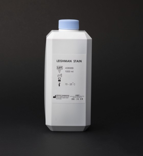
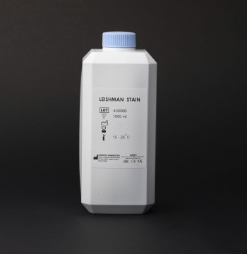
leishman stain
207.00
In Stock
Leishman stain is a type of histological stain primarily used in the study of blood smears and to examine the morphology of blood cells, as well as for detecting certain parasitic infections like Leishmaniasis. This stain is a modification of the Giemsa stain, and it is particularly useful for differentiating between different types of cells in blood and tissues.
Key Features of Leishman Stain:
✅ Composition: Leishman stain typically consists of eosin (which stains acidic structures) and methylene blue (which stains basic structures). These components work together to highlight different cellular components. ✅ Application: It's primarily used to stain blood smears, bone marrow samples, and tissue sections to identify various types of cells such as red blood cells, white blood cells, and platelets. It's also useful for detecting the presence of Plasmodium (the parasite that causes malaria) and Leishmania (the parasite responsible for Leishmaniasis). ✅ Ease of Use: It is easy to use and widely available in clinical laboratories for routine blood examination and parasitic identification.
Key Applications:
- Blood Smear Analysis: Leishman stain is used for examining the blood smear in diagnosing conditions like anemia, malaria, and various parasitic infections. It helps in identifying different types of white blood cells (e.g., neutrophils, lymphocytes, monocytes, etc.) and red blood cells.
- Leishmaniasis Diagnosis: The stain is used to detect Leishmania parasites in tissue samples, which are typically seen in the amastigote form.
- General Hematology: Used in routine hematological studies to examine blood cells for abnormalities, infections, or parasitic infections.
How Leishman Stain Works:
- Red Blood Cells: Stain pale pink or orange.
- White Blood Cells: The different types of white blood cells, such as neutrophils, lymphocytes, and monocytes, show different hues and intensities under the Leishman stain.
- Leishmania Parasites: These are typically identified within white blood cells, such as macrophages, in tissue smears.
Procedure for Using Leishman Stain:
Safety and Handling:
⚠️ Toxicity: Leishman stain contains chemicals that may be harmful if inhaled, ingested, or come into contact with skin. Handle with care, and wear appropriate protective equipment (e.g., gloves, lab coat, goggles).
⚠️ Proper Disposal: After use, dispose of the stained slides and chemicals according to laboratory safety protocols to avoid contamination or harmful exposure.
0 Reviews for leishman stain
Related Products
207.00
26.00
37.30

40.25

78.00
Customers Also Bought
9.20
234.00
63.25
40.25
158.07

46.00