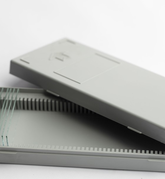
histology slides
105.80
Out of Stock
Histology Slides
Histology slides are thin sections of biological tissues that are stained and mounted on glass slides for microscopic examination. They are commonly used in medical, research, and educational settings to study tissue structure, function, and pathology.
Types of Histology Slides:
Normal Tissue Slides:
- Show the typical structure of organs and tissues.
- Used for teaching and comparative analysis.
Pathological Tissue Slides:
- Contain abnormal or diseased tissues.
- Used in medical diagnosis and research.
Special Stain Slides:
- Stained with different dyes to highlight specific structures (e.g., PAS, Masson's Trichrome, Silver Stains).
- Used to identify particular cell types, fibers, or microorganisms.
Frozen Section Slides:
- Rapidly prepared slides used in intraoperative pathology assessments.
Common Staining Techniques:
- Hematoxylin & Eosin (H&E): The most common stain, providing contrast between nuclei (blue) and cytoplasm (pink).
- Periodic Acid-Schiff (PAS): Highlights carbohydrates and glycogen.
- Masson's Trichrome: Differentiates muscle, collagen, and fibrin.
- Silver Stains: Used for nerve fibers and certain microorganisms.
- Immunohistochemistry (IHC): Uses antibodies to detect specific proteins in tissues.
Uses of Histology Slides:
???? Medical Diagnosis: Helps identify diseases like cancer, infections, and tissue abnormalities.
???? Research & Education: Used to study tissue structures and functions in academic settings.
???? Pharmaceutical Testing: Evaluates drug effects on tissues.
0 Reviews for histology slides
Related Products


126.05

51.75
138.00
79.35